
Methods
1. Immunohistochemistry
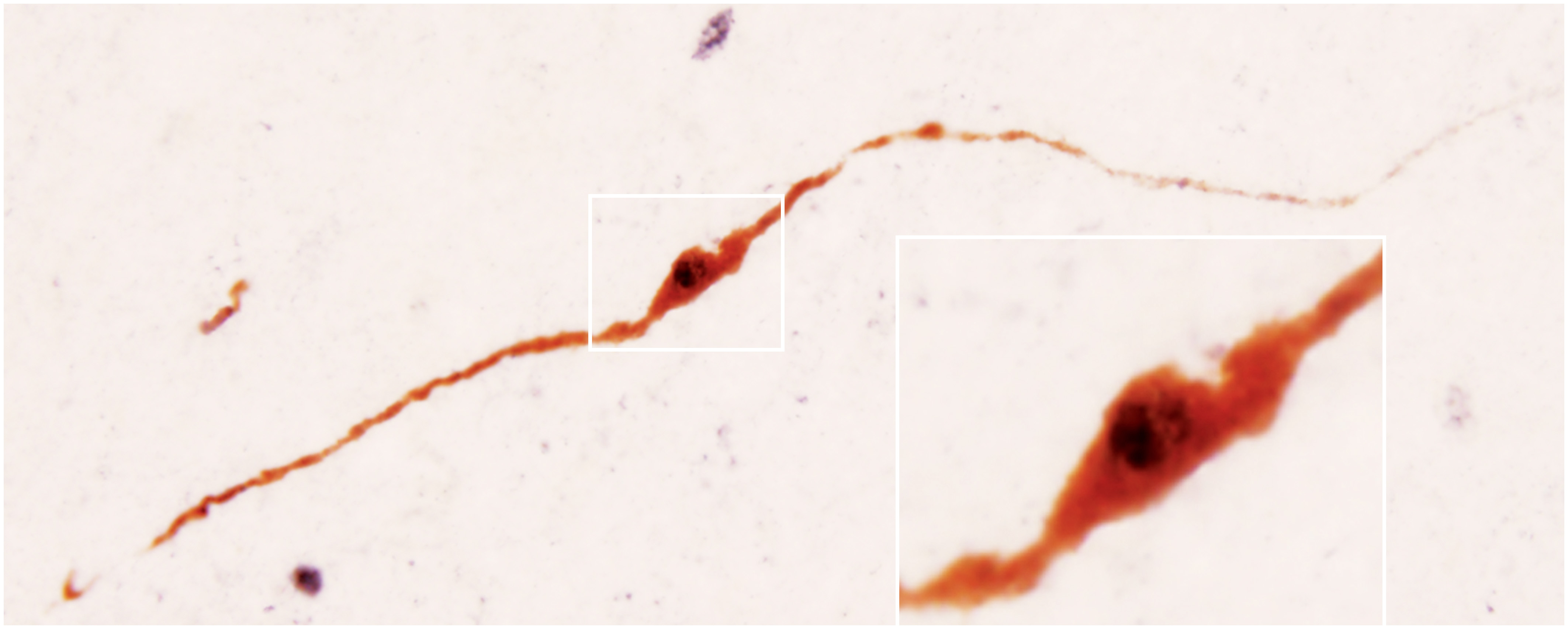 Both immersion-fixed and perfusion-fixed hypothalami are used routinely in immunohistochemical studies. The figure illustrates the results of double-label immunohistochemical experiments which revealed estrogen receptor beta immunoreactivity (black) in human GnRH neurons (brown).
2. Morphometric analysis of immuno-labeled hypothalamic neurons
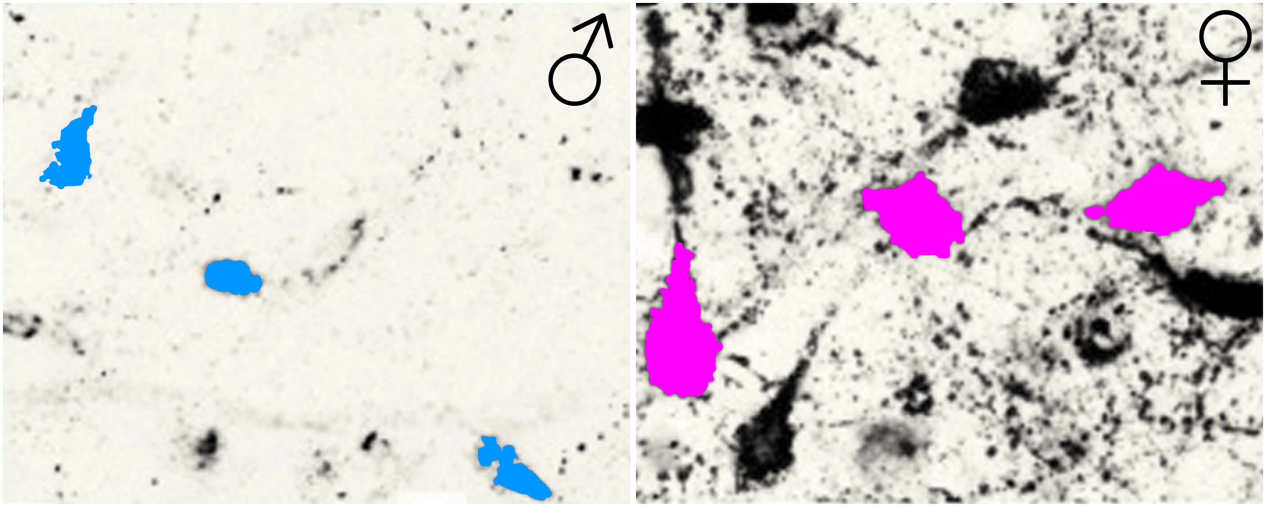 The large number of the immersion-fixed tissue specimens enables the morphometric studies of neurons to establish sex differences and aging-related changes. The example in the figure reveals a significant sex difference in the size (mean profile area) of human kisspeptin neurons in the infundibular nucleus of middle-aged male and female humans. The increased cell size in postmenopausal women is a consequence of a neuronal hypertrophy in the absence of estrogens (for details, see Hrabovszky et al., Front Endocrinol (Lausanne): 2:80).
3. Immunofluorescent multiple-labeling and confocal microscopy
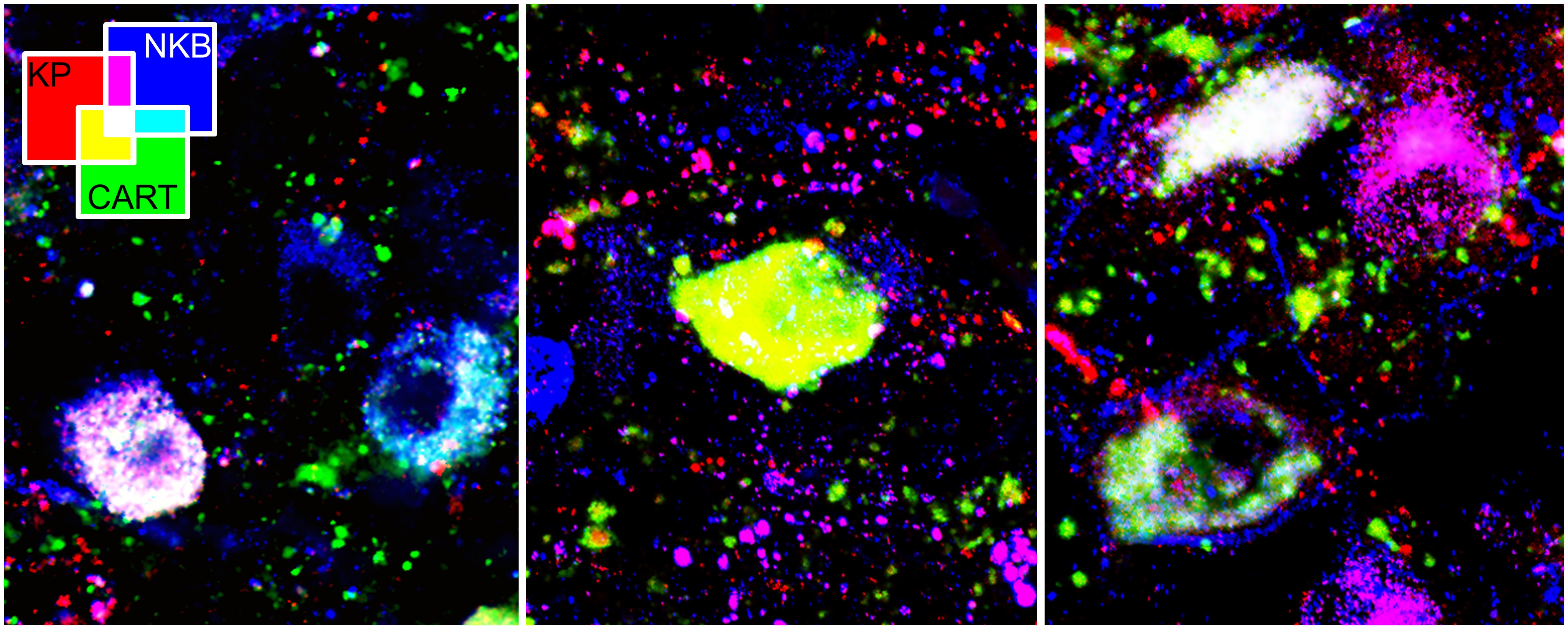 This picture illustrates the discovery of cocaine- and amphetamine-regulated transcript (CART) in a subset of kisspeptin (KP) and neurokinin B (NKB)-containing neurons of the human mediobasal hypothalamus (for details, see Skrapits et al., PLoS One: 9(8): e103977).
4. Quantitative immunohistochemistry
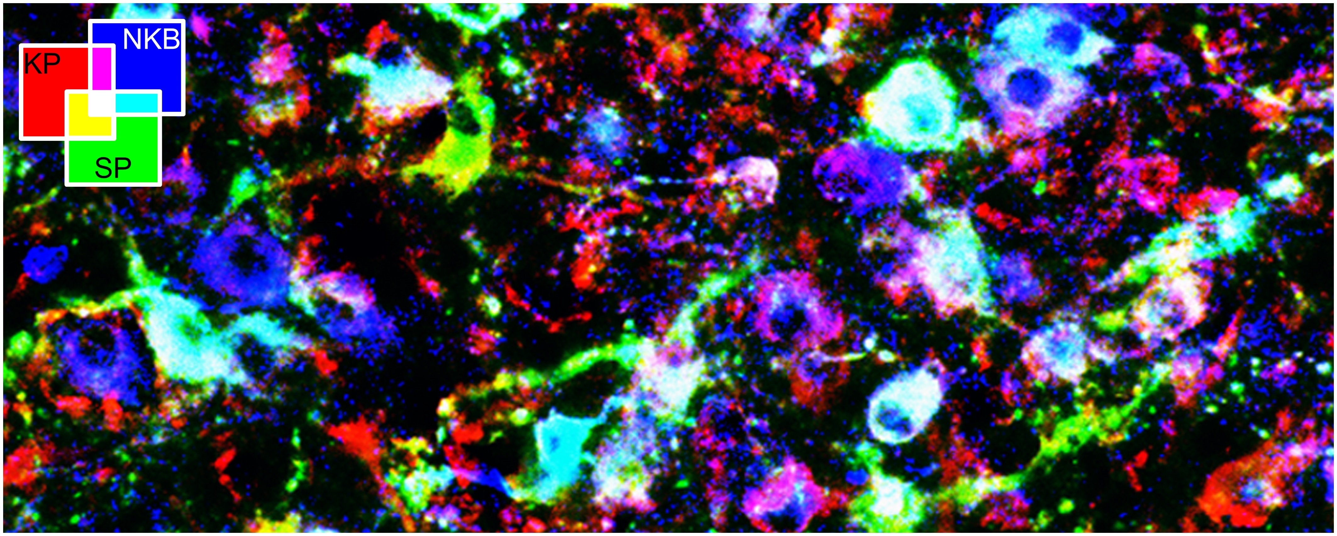 The immersion-fixed tissue samples are compatible with the quantitative analysis of neuropeptide coexpression patterns. This figure depicts the results of colocalization experiments between substance P (SP), kisspeptin (KP) and neurokinin B (NKB) immunofluorescent signals within neuronal cell bodies of the human infundibular nucleus (for details of this study, see Hrabovszky et al., PLoS One:8(8): e72369).
5. Preembedding immunoelectron microscopy
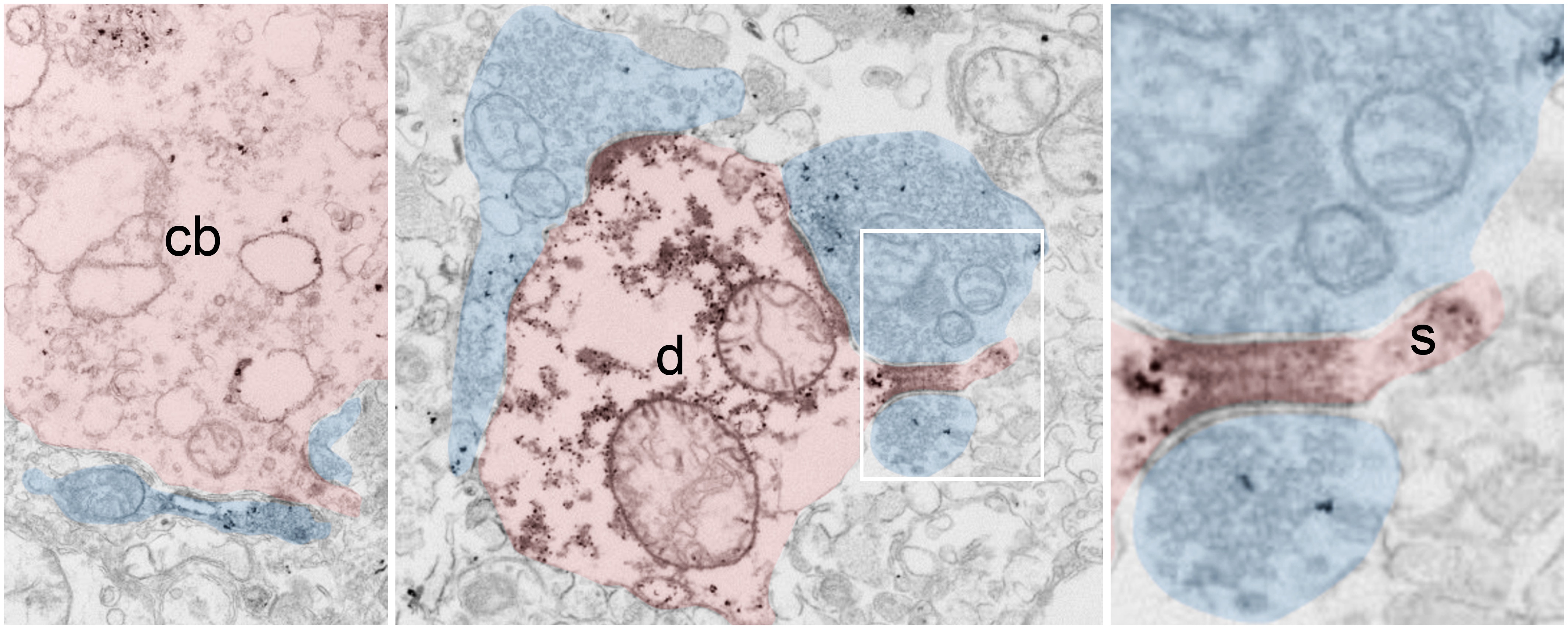 Short post mortem-time immersion-fixed hypothalami are compatible with immunoelectron microscopy. These figures reveal kisspeptin-immunoreactive axons (semi-transparent blue) establishing asymmetric synapses with kisspeptin-immunoreactive cell bodies (semi-transparent red, cb), dendrites (d) and dendritic spines (s) in the human infundibular nucleus (Takács et al., in press).
6. In situ hybridization with radioisotopic and digoxigenin-labeled cRNA probes
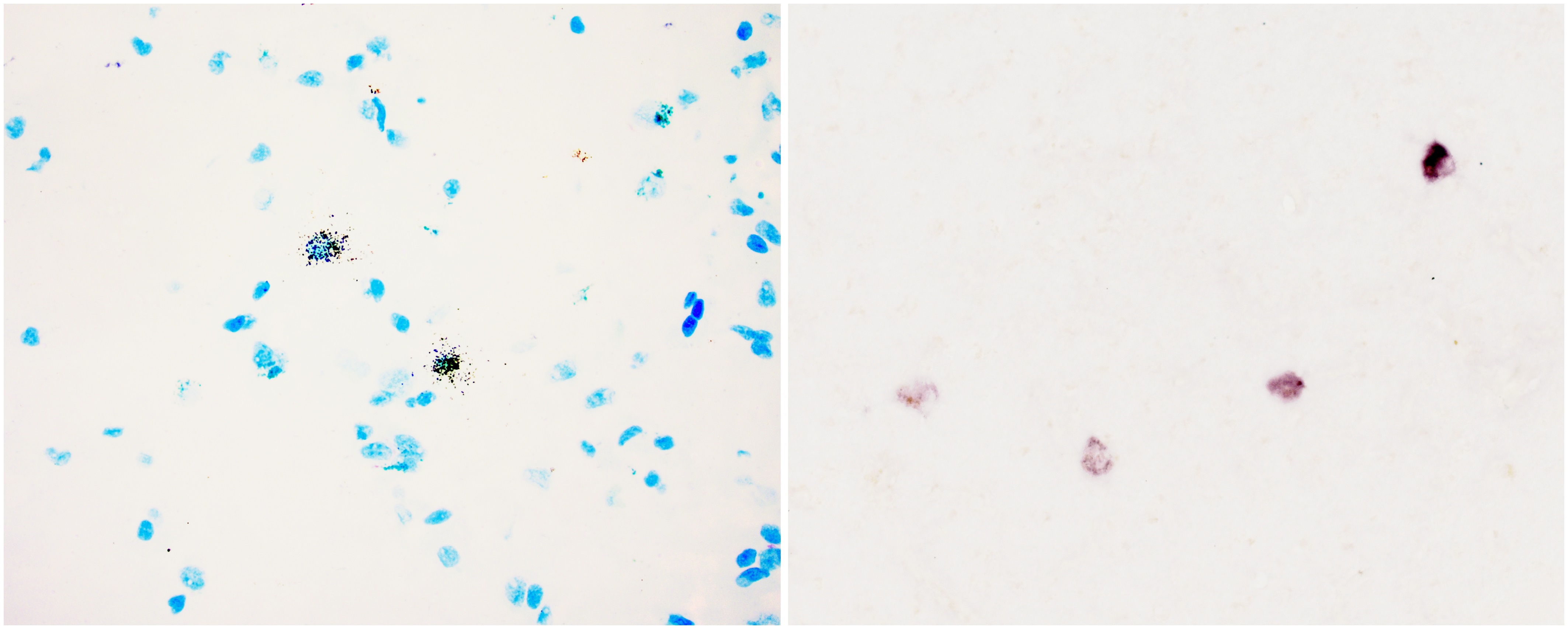 Short post mortem-time tissues are compatible with in situ hybridization experiments. The figures illustrate autoradiographic in situ hybridization signal to RF-amide related peptide mRNA in the hypothalamic periventricular nucleus (left) and non-isotopic in situ hybridization signal to proGnRH mRNA in the human putamen (right) (in preparation).
7. Random DiI-labeling and 3D-reconstruction of immunolabeled neurons in post mortem brains
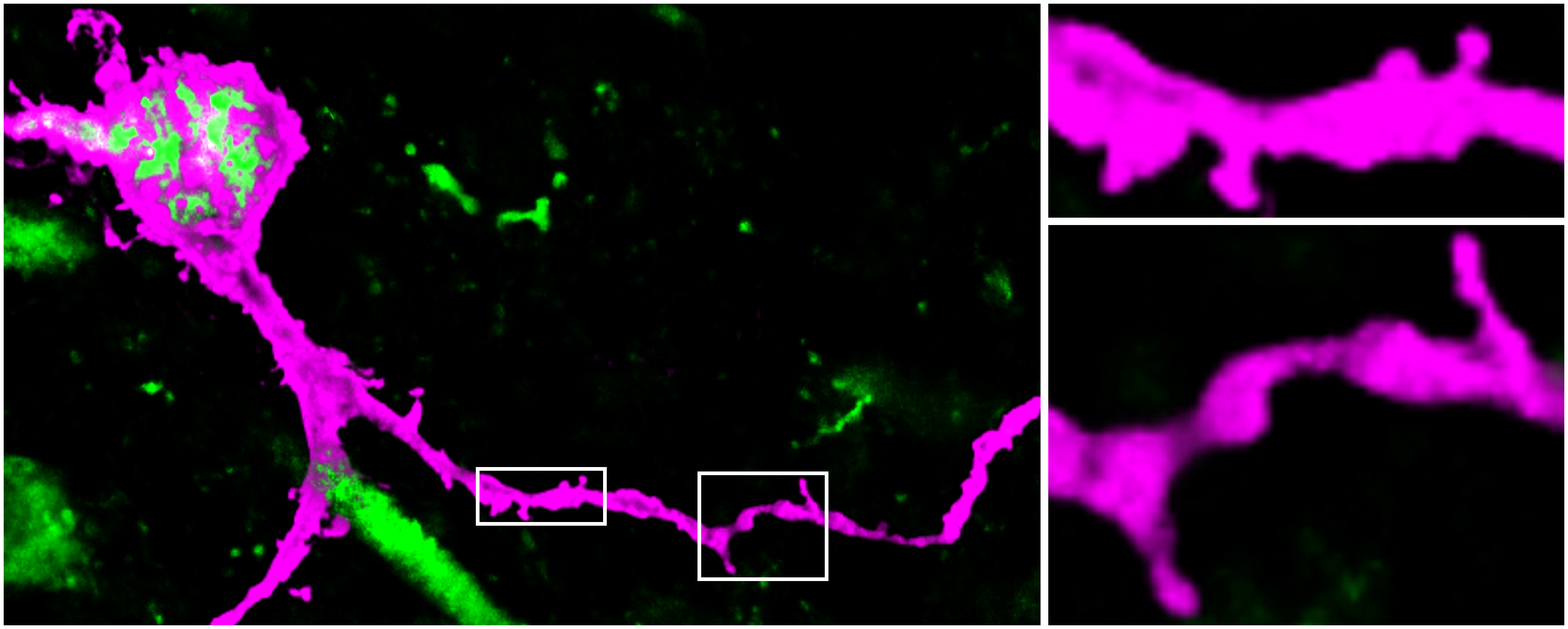 This figure illustrates the ‘diolistic’ labeling of an immunohistochemically identified kisspeptin neuron (green) using a Gene Gun loaded with the lipophilic dye DiI adsorbed to tungsten beads. The DiI signal (magenta) reveals the presence of numerous somatic and dendritic spines in double-labeled kisspeptin neurons (Takács et al., in press).
8. RT-qPCR studies of microdissected hypothalamic nuclei
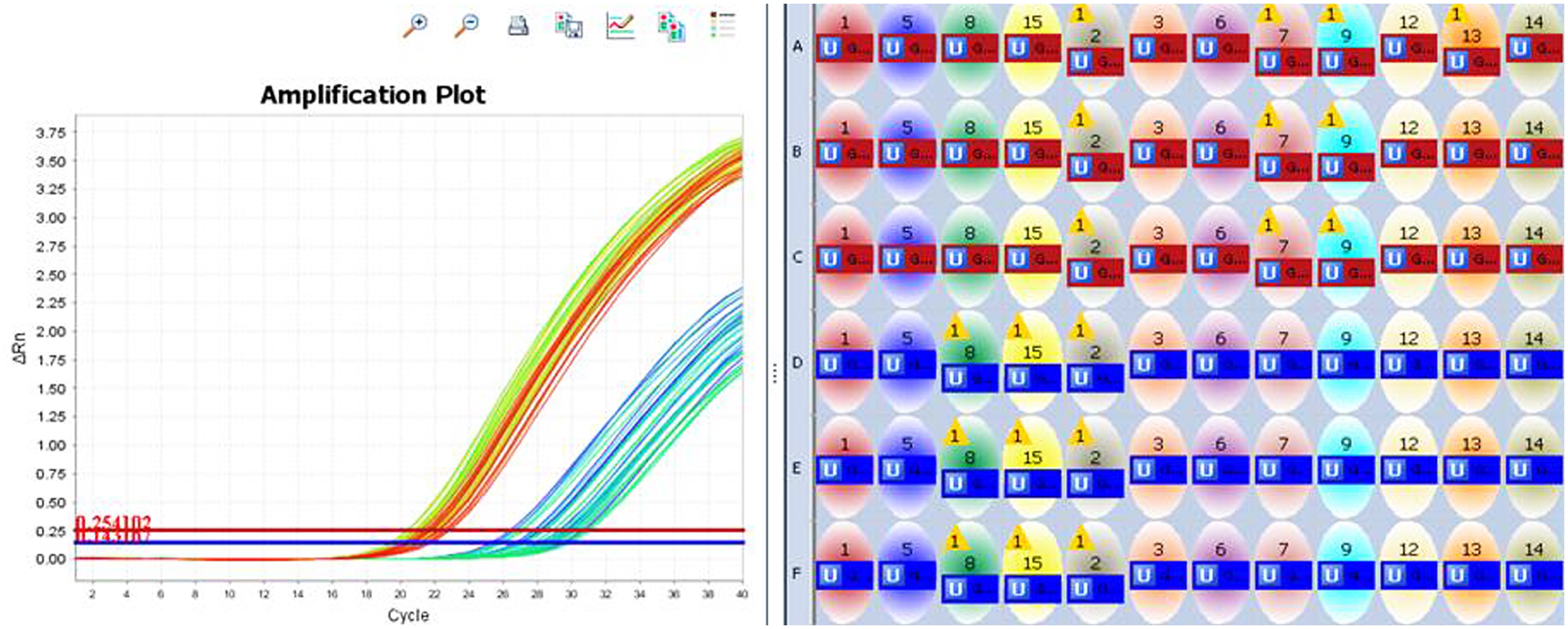 The figure demonstrates the successful use of micropunched hypothalamic nuclei for RT-qPCR amplification of VGLUT1 and VGLUT2 mRNAs in the human mediobasal hypothalamus (Sárvári et al., in preparation).